
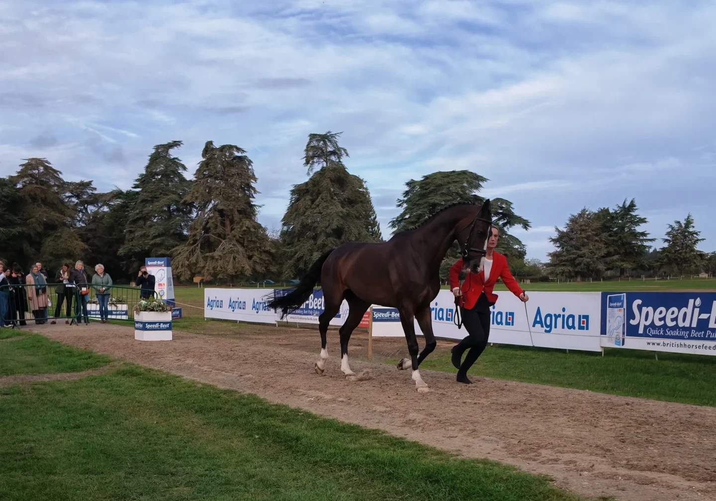
The superficial digital flexor tendon (SDFT) plays a crucial role in storing and releasing energy during movement and works close to its limits during strenuous exercise. Research suggests that this tendon develops and matures early in life, but its ability to adapt to continued workload is limited. As a result, the SDFT can undergo gradual degeneration over time, which may increase the risk of injury, particularly in very active horses. (1)
How does the Superficial Digital Flexor Tendon (SDFT) become injured?
The SDFT is particularly vulnerable because it is an energy-storing tendon. During fast work such as galloping and jumping, it stretches and recoils with every stride, storing and releasing energy to improve locomotor efficiency. During maximal exercise, the SDFT operates very close to its functional limits, making it susceptible to overload.
Main causes of SDFT injury
1. Repetitive overload rather than a single event
Most SDFT injuries develop from repeated loading cycles during training rather than a single traumatic incident. Small amounts of microscopic damage accumulate within the tendon matrix, and if the tendon is repeatedly stressed before it has repaired previous microdamage, this damage progressively accumulates until a clinical injury occurs.
2. Insufficient recovery between intense exercise sessions
Significant structural changes occur within the tendon after strenuous exercise. The research suggests the tendon may require up to 72 hours to return to normal homeostasis after intense loading. Training or galloping again before this recovery period can increase the chance of injury.
3. Mechanical strain exceeds the tendon’s repair capacity
The tendon reaches a “metabolic inflection point” when:
- Damage occurs faster than repair.
- Nutrient supply and cellular repair mechanisms cannot keep up with tissue breakdown.
This leads to irreversible structural damage.
4. Heat accumulation within the tendon
High-speed exercise generates substantial heat within the tendon where core SDFT temperatures may reach 43°C to 45°C, and higher temperatures are possible in elite racehorses. Temperatures above approximately 42.5°C can impair tendon cell (tenocyte) survival. Additionally, thermal damage can cause cell death, matrix degeneration, and predispose the tendon to rupture.
5. Training and environmental risk factors
- Excessive training intensity or frequency
- Rapid increases in workload
- Inadequate conditioning programmes
- Artificial or unsuitable training surfaces
- Repeated jumping and greater fence heights
- Uneven terrain and gradients
- Use of boots or bandages that increase heat retention
- Insufficient recovery periods
6. Age-related degeneration as horses age
- Elastin within the tendon decreases.
- The interfascicular matrix (IFM), which allows tendon fascicles to slide and recoil, becomes stiffer.
- Fatigue resistance decreases.
- Repair mechanisms become less effective.
Older horses therefore have a significantly greater risk of SDFT injury.
What happens when the SDFT is injured?
Initially, microscopic damage develops within the collagen matrix. As the damage accumulates, collagen fibres become disrupted and tendon cells release inflammatory and catabolic mediators.
Areas of cell death develop in the tendon core where characteristic “core lesions” form. Tendon elasticity and strength decrease, and the injured tendon becomes less capable of storing and returning energy.
The tendon will gradually change from a highly organised structure into tissue containing fragmented collagen fibres, disorganised matrix, reduced mechanical strength, and increased susceptibility to reinjury.
How does the SDFT recover?
Tendon healing occurs in three overlapping phases:
1. Inflammatory phase: This is the body’s immediate response to injury.
- Removal of damaged tissue.
- Influx of inflammatory cells.
- Release of growth factors.
- Initiation of repair mechanisms.
2. Proliferative phase: This restores basic continuity to the tendon but does not recreate normal tendon architecture.
- Fibroblasts produce a temporary repair matrix.
- Large amounts of Type III collagen are laid down.
- Scar tissue begins to bridge the damaged area.
3. Re-modelling phase: The repair tissue gradually matures.
- Type III collagen is replaced by more Type I collagen.
- Tensile strength improves.
- The scar contracts and re-organises.
However, the repaired tendon does not fully return to its original structure because adult tendons have limited regenerative capacity. Unlike bone or muscle, the SDFT generally heals through fibrosis (scar formation) rather than true regeneration. This causes the collagen fibres to remain less organised, and the normal interfascicular structure is not fully restored. As a result, the tendon’s energy-storing function is reduced.
Even when an ultrasound indicates healing, the tissue often remains mechanically inferior to the original tendon. This explains why reinjury rates remain high, and approximately 23% to 67% of horses suffer a repeat tendon injury within two years after conservative treatment.
Factors that support recovery include:
- Adequate recovery between intense exercise sessions (at least 72 hours between periods of heavy tendon loading)
- Progressive and evidence-based rehabilitation programmes
- Monitoring with ultrasound or ultrasound tissue characterisation (UTC)
- Early detection of inflammation through thermography and clinical assessment
- Management of tendon temperature after exercise
- Cryotherapy (cold therapy), which can:
- Rapidly reduce tendon core temperature
- Reduce metabolism and inflammatory responses
- Limit secondary tissue damage
- Low-impact exercise, such as water treadmills and aquatic conditioning, which may reduce tendon loading while maintaining fitness. (2)
Summary
The SDFT becomes injured when repeated mechanical and thermal stresses exceed the tendon’s ability to repair microscopic damage. Injury is usually a cumulative process, driven by overload, insufficient recovery, heat build-up and age-related degeneration.
Recovery occurs through inflammation, scar tissue formation and re-modelling, but the tendon rarely regenerates its original structure. As a result, healed tendons remain biomechanically weaker and are prone to reinjury, highlighting the importance of controlled rehabilitation, sufficient recovery periods and early detection of tendon stress. (2)
The use of the ArcEquine for superficial digital flexor tendon (SDFT):
When tissue is injured, the normal electrical activity of the cells is disrupted. The damaged area often develops a higher resistance to electrical signals than the surrounding healthy tissue, making it harder for the body’s natural bioelectrical currents to pass through the injury. This can slow down the normal healing processes.
Microcurrent therapy is thought to help by providing a gentle external electrical current that supports the body’s own natural electrical signals. By helping restore normal electrical activity in the injured tissue, microcurrent may reduce the resistance within the damaged area and improve communication between cells. This may encourage the tissue to return to a balanced, healthy state and support the body’s natural healing mechanisms. (3)
Protocol to follow:
Pre-2026 Delivery Unit:
Injury Protocol:
P1: daily for 1 week or until sound at walk (whichever is longer),
P2: daily for 2 weeks,
P3: daily for 3 weeks,
Once completed, use programme 2 or 4 (P4) for a minimum of two days per month.
2026 Delivery Unit:
Injury Protocol:
P1: daily for 1 week or until sound at walk (whichever is longer),
P2: daily for 2 weeks,
P3: daily for 3 weeks,
P4: daily for 4 weeks, or until healing is complete (whichever is longer)
Once completed, use programme 3 (P3) for a minimum of two days per month.
- Dowling BA, Dart AJ, Hodgson DR, Smith RKW. Superficial digital flexor tendonitis in the horse. Equine Vet J. 2000;32(5):369-378.
- O’Brien C, Marr N, Thorpe CT. Microdamage in the equine superficial digital flexor tendon. Equine Vet J [Internet]. 2021;53(3):417-430 [cited 2026 Sep 29]. Available from: https://beva.onlinelibrary.wiley.com/doi/full/10.1111/evj.13331
- Lin YL, Moolenaar H, van Weeren PR. Effect of microcurrent electrical tissue stimulation on equine tenocytes in culture. Am J Vet Res. 2006;67(2):271-276.